ECGs by diagnosis: Difference between revisions
Neil.m.young (talk | contribs) (link added) |
|||
| Line 34: | Line 34: | ||
==Hypothermia== | ==Hypothermia== | ||
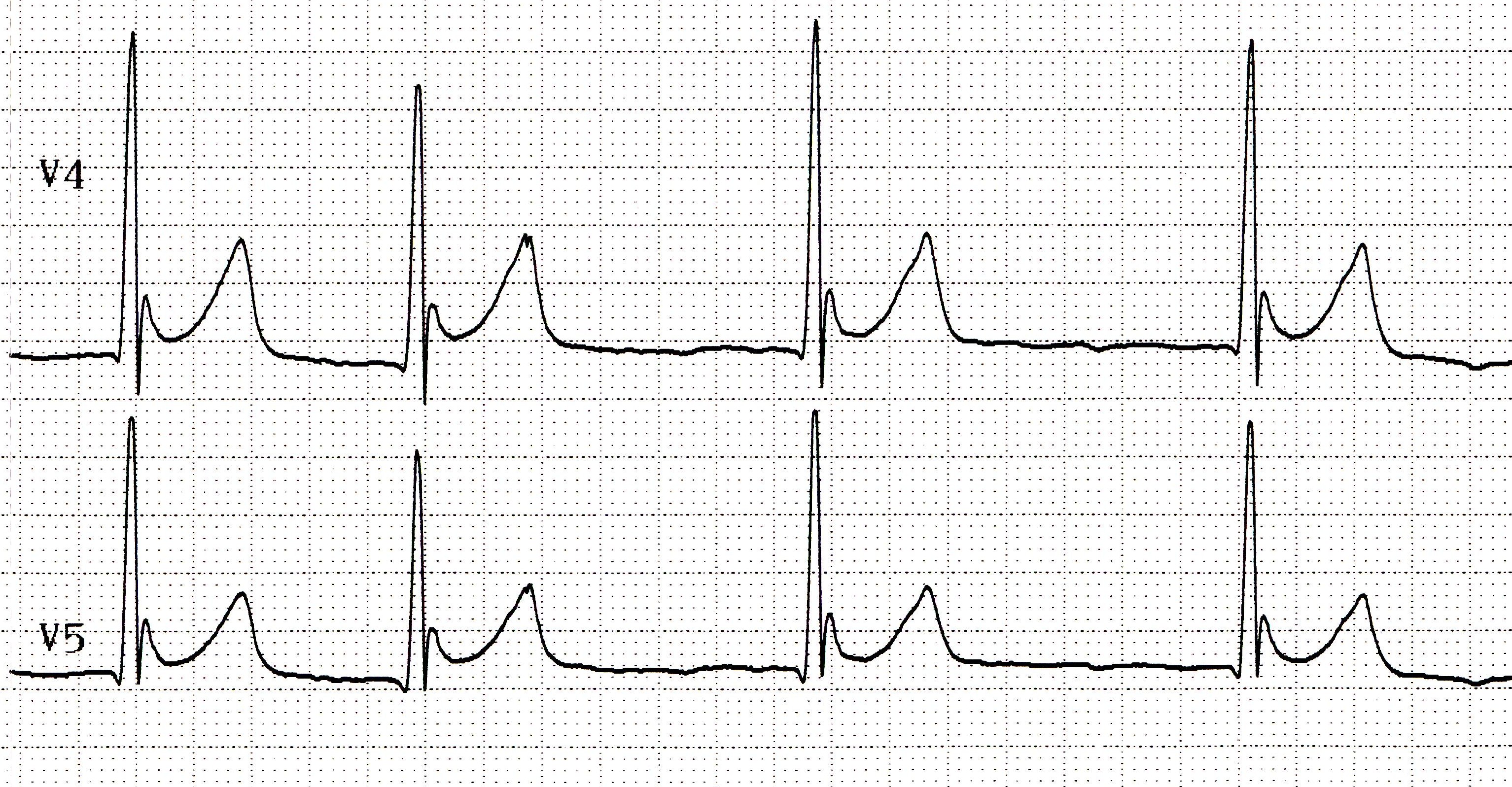
#Osborn wave (J wave) - Positive deflection at the J point | #Osborn wave (J wave) - Positive deflection at the J point | ||
##height of the J wave correlates to the degree of hypothermia [[Media:jwaves.jpg]] | |||
#Bradyarrhythmias, AV blocks | #Bradyarrhythmias, AV blocks | ||
#Prolonged PR, QRS, QT intervals | #Prolonged PR, QRS, QT intervals | ||
Revision as of 03:04, 1 December 2015
ACS
- See ACS
Aneurysm
- Suggested by:
- ST elevation >4wk
- QS wave in setting of ST-segment elevation w/o T-wave inversion
Pericarditis
- See Pericarditis
Electrolyte Disorders
CNS
- SAH, IC Bleed, CVA
- Diffuse wide, deep, blunted, inverted T waves
- QT Prolongation
Pacemakers
- Should be in the apex of R ventricle
- ECG should mimic LBBB w/ LAD
Pulmonary Embolism
- Sinus tachycardia
- S1Q3T3 (Sp, not Sn)
- Right axis deviation
- RBBB
- T wave inversions leads V1-V3
Hypothermia
- Osborn wave (J wave) - Positive deflection at the J point
- height of the J wave correlates to the degree of hypothermia Media:jwaves.jpg
- Bradyarrhythmias, AV blocks
- Prolonged PR, QRS, QT intervals
- Shivering artifact