Epidural hemorrhage: Difference between revisions
No edit summary |
No edit summary |
||
| Line 24: | Line 24: | ||
==Management== | ==Management== | ||
* Emergent neurosurgical evacuation | *Emergent neurosurgical evacuation | ||
* Bilateral trephination (burr holes) if neurosurgery is unavailable | *Bilateral trephination (burr holes) if neurosurgery is unavailable | ||
*Medical care - general goal of decreasing ICP<ref>Price DD, et al. Epidural Hematoma in Emergency Medicine Treatment and Management. Updated Dec 9, 2014. http://emedicine.medscape.com/article/824029-treatment#a1126</ref> | |||
**RSI with possible lidocaine and fentanyl premedication | |||
**Elevate HOB 30 degrees (or reverse Trendelenburg position) | |||
**If continued signs of increasing ICP: | |||
***Mannitol 0.25 - 1 g/kg IV if MAP > 90 mmHg after NSGY c/s | |||
***Hyperventilation to 30-35 mmHg, no lower than 25 mmHg | |||
==Disposition== | ==Disposition== | ||
Revision as of 16:39, 11 June 2015
Background
- Occur as a result of blood collecting between the skull and the dura mater
- Most commonly secondary to a tear of the middle meningeal artery
Clinical Features
- Generally associated with blunt trauma to the temporal or temporoparietal region
- There is a high incidence of associated skull fractures (>75%) and additional cerebral injuries (intraparenchymal hemorrhage, cerebral contusion, contrecoup injuries, subdural hematoma, subarachnoid hemorrhage)
Differential Diagnosis
Intracranial Hemorrhage Types
- Intra-axial
- Hemorrhagic stroke (Spontaneous intracerebral hemorrhage)
- Traumatic intracerebral hemorrhage
- Extra-axial
- Epidural hemorrhage
- Subdural hemorrhage
- Subarachnoid hemorrhage (aneurysmal intracranial hemorrhage)
Diagnosis
- Any patient with a neurologic deficit, depressed GCS, palpable skull fracture, or worrisome mechanism will warrant a non-contrast head CT after initial stabilization and resuscitation.
- Canadian CT Head Rule for patients with minor head injury
- Can be used to decide which minor injuries will require head CT
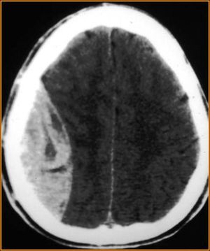
- Findings on CT are, classically, a lens (or lemon-shaped) shaped hyperdense lesion with sharp margins in the temporoparietal region
- Blood along the inside of the skull will not cross the sutures. This helps differentiate acute epidural hematoma from acute subdural hematoma.
Workup
Workup
- Consider head CT (rule out intracranial hemorrhage)
- Use validated decision rule to determine need
- Avoid CT in patients with minor head injury who are at low risk based on validated decision rules.[1]
- Consider cervical and/or facial CT
- Appropriate trauma resuscitation of all patients with head trauma
- A thorough neurological examination of any patient with head trauma BEFORE administration of RSI
Management
- Emergent neurosurgical evacuation
- Bilateral trephination (burr holes) if neurosurgery is unavailable
- Medical care - general goal of decreasing ICP[2]
- RSI with possible lidocaine and fentanyl premedication
- Elevate HOB 30 degrees (or reverse Trendelenburg position)
- If continued signs of increasing ICP:
- Mannitol 0.25 - 1 g/kg IV if MAP > 90 mmHg after NSGY c/s
- Hyperventilation to 30-35 mmHg, no lower than 25 mmHg
Disposition
- Transfer to tertiary medical center
- Admission to NS or Trauma Surgery
See Also
External Links
References
- Stiell IG, Wells GA, Vandemheen K, et al. The Canadian CT Head Rule for patients with minor head injury. Lancet. 2001;357(9266):1391-6.
- Judith E. Tintinalli, Gabor Kelen, J. Stephan Stapczynski. SAMJ. New York : McGraw-Hill, Medical Pub. Division, c2004.; 2008.
- Irie F, Le Brocque R, Kenardy J et-al. Epidemiology of traumatic epidural hematoma in young age. J Trauma. 2011;71 (4): 847-53.
- ↑ Choosing wisely ACEP
- ↑ Price DD, et al. Epidural Hematoma in Emergency Medicine Treatment and Management. Updated Dec 9, 2014. http://emedicine.medscape.com/article/824029-treatment#a1126


