Indications
- Suspicion of septic arthritis
- Suspicion of crystal induced arthritis
- Evaluation of therapeutic response for septic arthritis
- Unexplained arthritis with synovial effusion
Relative Indications
- Therapeutic (decrease intra-articular pressure, injection of anesthetics/steroids)
Contraindications
- No absolute contraindications for diagnostic arthrocentesis
- Do not inject steroids into a joint that you suspect is already infected
- Relative Contraindications:
- Overlying cellulitis
- Coagulopathy
- Joint prosthesis (refer to ortho)
Equipment Needed
- Betadine or Chlorhexadine
- Sterile gloves/drape
- Sterile gauze
- Lidocaine
- Syringes
- Small syringe (6-12cc) for injection of local anesthetic
- Large syringe (one 60cc or two 30cc) for aspiration
- Needles
- 18 gauge
- 27 gauge
- Collection tubes (red top)
- Culture bottles
Procedure
- Prep area w/ betadine or chlorhexadine using circular motion moving away from joint x 3
- Drape joint in sterile fashion
- Inject lidocaine w/ 25-30ga needle superficially and then into deeper tissues
- Insert 18ga needle (for larger joints) into joint space while pulling back on syringe #Stop once you aspirate fluid; aspirate as much fluid as possible
- Send: cell count, culture, Gram stain, crystal analysis
Approach
Shoulder
- Anterior approach
- Sit pt upright facing you
- Insert needle just lateral to coracoid process (between coracoid process and humeral head)
- Direct needle posteriorly
- Posterior Approach
- Sit pt upright w/ back facing you
- Palpate scapular spine to its lateral limit (the acromion)
- Identify the posterolateral corner of the acromion
- Insert 1.5in needle 1 cm inferior and 1 cm medial to this corner
- Direct needle anterior and medial toward presumed position of coracoid process
- Glenohumeral joint is located at a depth of approximately 1-1.5in
Elbow
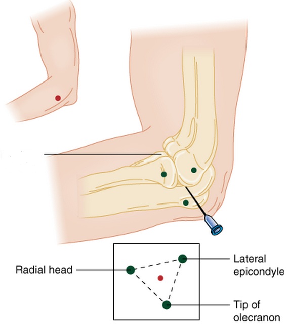
- Place elbow in 90' flexion, resting on a table, w/ hand prone
- Locate radial head, lateral epicondyle , and lateral aspect of olecranon tip
- These landmarks form the anconeus triangle
- Palpate a sulcus just proximal to the radial head (in the middle of the triangle)
- Insert needle into sulcus directed medial and perpendicular to radius toward distal end of antecubital fossa

Wrist
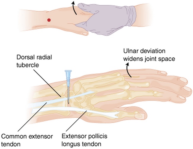
- Palpate landmarks w/ wrist in neutral position:
- Radial tubercle of distal radius
- Anatomic snuffbox
- Extensor pollicis longus tendon
- Common extensor tendon of index finger
- Insert needle perpendicular to skin, ulnar to radial tubercle and anatomic snuffbox, between extensor pollicis longus and common extensor tendons

Knee
- Can be entered medially or laterally to the patella
- Fully extend knee and ensure quadriceps muscle is relaxed
- Identify midpoint of patella; insert needle either lateral or medial
- Direct needle posterior to patella and horizontally toward the joint space
- Compression or "milking" applied to both sides of joint space may facilitate aspiration
Ankle
- Lateral approach (subtalar)
- Keep foot perpendicular to leg
- Enter subtalar joint just below tip of lateral malleolus
- Direct needle medially toward joint space
- Medial approach (tibiotalar)
- Have pt supine w/ foot perpendicular to leg
- Palpate sulcus lateral to medial malleolus and medial to TA and EHL tendons
- Then plantarflex foot w/ needle entering skin overlying the sulcus
- Angle needle slightly cephalad as it passes between medial malleolus and TA tendon
Metacarpophalangeal
- have palm facing down and apply gentle traction to the affected digit
- insert needle dorsally just medial or lateral to midline and proximal to the base of the proximal phalanx
Interphalangeal
- have palm facing down and apply gentle traction to the affected digit
- insert needle dorsally medial or lateral to midline and proximal to base of middle or distal phalanx
Metatarsophalangeal
- patient supine with flexion of the MTP joint 15-20 degrees and apply gentle traction
- insert needle dorsally just medial or lateral to midline between the metatarsal head and base of proximal phalanx
Interphalangeal
- patient supine with joint flexed 15-20 degrees with gentle traction
- insert needle dorsally, medial or lateral to midline between head of proximal phalanx and base of more distal phalanx
Complications
- pain
- infection
- reaccumulation of effusion
- damage to tendons, nerves, or blood vessels
See Also
Septic Arthritis (General)
Monoarticular Arthritis
Septic Arthritis (Hip)
Septic Arthritis (Peds)
Source


