Thoracic and lumbar fractures and dislocations: Difference between revisions
No edit summary |
|||
| (50 intermediate revisions by 10 users not shown) | |||
| Line 1: | Line 1: | ||
== | ==Background== | ||
*Injury to thoracic spine necessitates severe force | *Injury to thoracic spine necessitates severe force | ||
**thoracic spine has enhanced stiffness secondary to articulations with the rib cage | |||
**When spinal cord injury occurs usually complete | **When spinal cord injury occurs usually complete | ||
*Stable if two or more of the spinal columns are intact: | **thoracic spinal canal is narrower than in other regions, increased risk of cord injury | ||
*Important to evaluate for thoracic spine injuries and aortic injuries in the setting of blunt chest trauma with mediastinlal widening | |||
*Follows the three column model - [[Unstable spine fractures|Stable]] if two or more of the spinal columns are intact: | |||
**Anterior (anterior longitudinal ligament, annulus fibrosus, ant. half of the vertebral body) | **Anterior (anterior longitudinal ligament, annulus fibrosus, ant. half of the vertebral body) | ||
**Middle (posterior longitudinal ligament, posterior annulus fibrous, and post. half of vertebral body | **Middle (posterior longitudinal ligament, posterior annulus fibrous, and post. half of vertebral body | ||
**Posterior (supraspinous and interspinous ligaments, facet joint capsule) | **Posterior (supraspinous and interspinous ligaments, ligamentum flavum, facet joint capsule) | ||
*Unstable if: | *Unstable if: | ||
**50% loss of vertebral height | **50% loss of vertebral height | ||
**Kyphotic angulation around the | **Kyphotic angulation around the fracture: | ||
***> | ***>30' for compression fracture | ||
***> | ***> 25' for burst fracture | ||
**Neurologic deficit | **Neurologic deficit | ||
{{Vertebral fractures and dislocations types}} | |||
== | ==Clinical Features== | ||
* | *Typically pain over site of injury | ||
=== | ==Differential Diagnosis== | ||
{{Thoracic trauma DDX}} | |||
{{Lower back pain DDX}} | |||
=== | ==Evaluation== | ||
* | [[File:T12compressionfracMark.png|thumb|[[Thoracic compression fracture]] of T12.]] | ||
* | ===Workup=== | ||
** | *Type and screen/cross, labs including pancreatic enzymes if thoraco-lumbar location | ||
*** | |||
* | *Indications to Image Thoracic and Lumbar Spine after Trauma | ||
** Compression | **Mechanism | ||
***Gunshot, High energy trauma, Motor vehicle crash with rollover or ejection, Fall >10 ft or 3 m, Pedestrian hit by car | |||
**Physical Exam | |||
***Midline back pain, Midline focal tenderness, Evidence of spinal cord or nerve root deficit | |||
**Associated injuries | |||
***Cervical fracture, ribe fracture, aortic injuries, hollow viscus injuries | |||
*Plain radiographs or CT scan to evaluate for body abnormality | |||
*Can reformat Chest and Abdomen CT to look at thoracic, lumbar spine | |||
*MRI is diagnostic test of choice to evaluate patients with nerve injury | |||
*CT myelography alternative when MRI unavailable | |||
*anterior vertebral body compression fracture with extension through middle of vertebral body into posterior wall | |||
*Compression fracture + increased posterior interspinous spaces caused by distraction | |||
10% of patients with a spine fracture have second fracture in a different segment | |||
CT IF: | |||
*Compression | |||
*Wedge | |||
*>50% height (rule out middle column & burst) | |||
===Diagnosis=== | |||
==Management== | |||
*Spinal precautions | |||
*Consult ortho or neurosurgery (institution dependent) | |||
*Stable fractures | |||
**TLSO brace in discussion with consulting service | |||
*Unstable fractures | |||
**Emergency operative repair unless medically unstable | |||
==Disposition== | |||
==See Also== | ==See Also== | ||
[[Spinal Cord Trauma]] | *[[Spinal Cord Trauma]] | ||
*[[Vertebral fractures]] | |||
==External Links== | |||
*[https://www.east.org/education/practice-management-guidelines/thoracolumbar-spinal-injuries-in-blunt-trauma%2C-screening-for EAST Guidelines for screening for thoracolumbar injuries] | |||
== | ==References== | ||
<references/> | |||
[[Category:Trauma]] | [[Category:Trauma]] | ||
[[Category:Neurology]] | |||
[[Category:Orthopedics]] | |||
Latest revision as of 17:13, 27 October 2020
Background
- Injury to thoracic spine necessitates severe force
- thoracic spine has enhanced stiffness secondary to articulations with the rib cage
- When spinal cord injury occurs usually complete
- thoracic spinal canal is narrower than in other regions, increased risk of cord injury
- Important to evaluate for thoracic spine injuries and aortic injuries in the setting of blunt chest trauma with mediastinlal widening
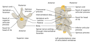
- Follows the three column model - Stable if two or more of the spinal columns are intact:
- Anterior (anterior longitudinal ligament, annulus fibrosus, ant. half of the vertebral body)
- Middle (posterior longitudinal ligament, posterior annulus fibrous, and post. half of vertebral body
- Posterior (supraspinous and interspinous ligaments, ligamentum flavum, facet joint capsule)
- Unstable if:
- 50% loss of vertebral height
- Kyphotic angulation around the fracture:
- >30' for compression fracture
- > 25' for burst fracture
- Neurologic deficit
Vertebral fractures and dislocations types
- Cervical fractures and dislocations
- Thoracic and lumbar fractures and dislocations
Clinical Features
- Typically pain over site of injury
Differential Diagnosis
Thoracic Trauma
- Airway/Pulmonary
- Cardiac/Vascular
- Musculoskeletal
- Other
Lower Back Pain
- Spine related
- Acute ligamentous injury
- Acute muscle strain
- Disk herniation (Sciatica)
- Degenerative joint disease
- Spondylolithesis
- Epidural compression syndromes
- Thoracic and lumbar fractures and dislocations
- Cancer metastasis
- Spinal stenosis
- Transverse myelitis
- Vertebral osteomyelitis
- Ankylosing spondylitis
- Spondylolisthesis
- Discitis
- Spinal Infarct
- Renal disease
- Intra-abdominal
- Abdominal aortic aneurysm
- Ulcer perforation
- Retrocecal appendicitis
- Large bowel obstruction
- Pancreatitis
- Pelvic disease
- Other
Evaluation

Thoracic compression fracture of T12.
Workup
- Type and screen/cross, labs including pancreatic enzymes if thoraco-lumbar location
- Indications to Image Thoracic and Lumbar Spine after Trauma
- Mechanism
- Gunshot, High energy trauma, Motor vehicle crash with rollover or ejection, Fall >10 ft or 3 m, Pedestrian hit by car
- Physical Exam
- Midline back pain, Midline focal tenderness, Evidence of spinal cord or nerve root deficit
- Associated injuries
- Cervical fracture, ribe fracture, aortic injuries, hollow viscus injuries
- Mechanism
- Plain radiographs or CT scan to evaluate for body abnormality
- Can reformat Chest and Abdomen CT to look at thoracic, lumbar spine
- MRI is diagnostic test of choice to evaluate patients with nerve injury
- CT myelography alternative when MRI unavailable
- anterior vertebral body compression fracture with extension through middle of vertebral body into posterior wall
- Compression fracture + increased posterior interspinous spaces caused by distraction
10% of patients with a spine fracture have second fracture in a different segment
CT IF:
- Compression
- Wedge
- >50% height (rule out middle column & burst)
Diagnosis
Management
- Spinal precautions
- Consult ortho or neurosurgery (institution dependent)
- Stable fractures
- TLSO brace in discussion with consulting service
- Unstable fractures
- Emergency operative repair unless medically unstable


