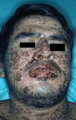
Exfoliative erythroderma
(Redirected from Erythroderma)
Background
- Also known as exfoliative dermatitis
- Diffuse, widespread scaly dermatitis that covers most of body surface
- Cutaneous reaction to a drug or chemical agent or underlying systemic or cutaneous disease
- Males affected twice as often as females
- Most patients >40 years old
Clinical Features
- Generalized erythema, warmth, scaling
- Can be pruritic and painful
- Abrupt onset if related to drug, contact allergen, or malignancy; gradual onset if related to underlying cutaneous disorder
- Generally starts on face and trunk with progression to other skin surfaces
- Low-grade fever common
- Tends to be a chronic condition, mean duration 5 years
Differential Diagnosis
- Erythema multiforme
- Stevens-Johnson syndrome and toxic epidermal necrolysis
- Drug rash
- DRESS syndrome
Erythematous rash
- Positive Nikolsky’s sign
- Febrile
- Staphylococcal scalded skin syndrome (children)
- Toxic epidermal necrolysis/SJS (adults)
- Afebrile
- Febrile
- Negative Nikolsky’s sign
- Febrile
- Afebrile
Evaluation
Workup
- CBC, CMP, ESR
- CXR
- Determine underlying cause, including evaluation for underlying malignancy and biopsy of skin
Diagnosis
Table of Severe Drug Rashes
| Charateristic | DRESS | SJS/TEN | AGEP | Erythroderma |
| Image |  |
 |
 |

|
| Onset of eruption | 2-6 weeks | 1-3 weeks | 48 hours | 1-3 weeks |
| Duration of eruption (weeks) | Several | 1-3 | <1 | Several |
| Fever | +++ | +++ | +++ | +++ |
| Mucocutaneous features | Facial edema, morbilliform eruption, pustules, exfoliative dermattiis, tense bullae, possible target lesions | Bullae, atypical target lesions, mucocutaneous erosions | Facial edema, pustules, tense bullae, possible target lesions, possibl emucosal involvement | Erythematous plaques and edema affecting >90% of total skin surface with or without diffuse exfoliation |
| Lymph node enlargement | +++ | - | + | + |
| Neutrophils | Elevated | Decreased | Very elevated | Elevated |
| Eosinophils | Very elevated | No change | Elevated | Elevated |
| Atypical lymphocytes | + | - | - | + |
| Hepatitis | +++ | ++ | ++ | - |
| Other organ involvement | Interstitial nephritis, pneumonitis, myocarditis, and thydoiditis | Tubular nephritis and tracheobronical necrosis | Possible | Possible |
| Histological pattern of skin | Perivascular lymphocytcic infiltrate | Epidermal necrosis | Subcorneal pustules | Nonspecific, unless reflecting Sezary syndrome or other lymphoma |
| Lymph node histology | Lymphoid hyperplasia | - | - | No, unless reflecting Sezary syndrome or other malignancy |
| Mortality (%) | 10 | 5-35 | 5 | 5-15 |
Management
- Emergent dermatology consult
- Fluid replacement for hypovolemia, monitor fluid intake
- Warming measures for hypothermia
- Wound care
- Discontinue all unnecessary medications
- Systemic corticosteroids after dermatology consult
- Antibiotics if evidence of secondary infection
Disposition
- Admit
Complications
- Hypothermia
- fluid/electrolyte/protein loss
- Invasion of bacteria and opportunistic organisms through the skin
- High-output heart failure due to vasodilatation
See Also
External Links
References
- Tintinalli's Emergency Medicine 7th Edition, pg 1614, 1617
- Harwood-Nuss' Clinical Practice of Emergency Medicine 6th Edition, pg 821


