Osteoarthritis
Background
- Osteoarthritis is a chronic arthropathy characterized by degeneration of joint cartilage and underlying bone
- Alternatively called arthrosis[1]
- Most common progressive joint disease
- Joints in the hands, knees, hips, and spine are the most frequently affected.
Risk Factors
- Age (almost exclusively in elderly)
- Female versus male sex
- Obesity
- Lack of osteoporosis
- Occupation
- Previous injury
- Muscle weakness
- Genetic elements
Clinical Features
- Commonly affected joints
- Cervical and lumbar spine
- First carpometacarpal joint
- Proximal interphalangeal joint
- Distal interphalangeal joint
- Hip
- Knee
- Subtalar joint
- First metatarsophalangeal joint
- Uncommonly affected joints
- Shoulder
- Wrist
- Elbow
- Metacarpophalangeal joint
Differential Diagnosis
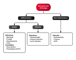
Monoarticular arthritis
- Acute osteoarthritis
- Avascular necrosis
- Crystal-induced (Gout, Pseudogout)
- Gonococcal arthritis, arthritis-dermatitis syndrome
- Nongonococcal septic arthritis
- Lyme disease
- Malignancy (metastases, osteochondroma, osteoid osteoma)
- Reactive poststreptococcal arthritis
- Trauma-induced arthritis
- Fracture
- Ligamentous injury
- Overuse
- Avascular necrosis
- Decompression sickness
- Spontaneous osteonecrosis
- Hemorrhagic (e.g. hemophilia, systemic anticoagulation
- Seronegative spondyloarthropathies (ankylosing spondylitis, IBD, psoriatic arthritis, reactive arthritis
- RA, SLE
- Sarcoidosis, amyloidosis
- Periarticular pathology
- Transient (Toxic) Synovitis (Hip)
- Slipped Capital Femoral Epiphysis (SCFE)
- Legg Calve Perthes Disease
Polyarthritis
- Fibromyalgia
- Juvenile idiopathic arthritis
- Lyme disease
- Osteoarthritis
- Psoriatic arthritis
- Reactive poststreptococcal arthritis
- Rheumatoid arthritis
- Rheumatic fever
- Serum sickness
- Systemic lupus erythematosus
- Serum sickness–like reactions
- Viral arthritis
Evaluation
- Greater than 50 years of age
- Morning stiffness for less than 30 minutes
- Crepitus on active motion of the knee
- Bony tenderness
- Bony enlargement
- No palpable warmth
Management
Nonpharmacological
- Weight loss
- Ice/warm packs
- Physical therapy
- Occupational therapy
- Exercise
Pharmacological [2]
AAOS Decision Tool for Knee OA
- Refer to this helpful tool from the American Academy of Orthopedic Surgeons for management and disposition options
- Link to knee osteoarthritis tool
Surgical
- Arthroscopy for removal of meniscal tears or loose foreign bodies
- No additional benefit compared to PT and medical therapy in routine OA [3]
- Osteotomy in young patients with misalignment of knee or hip joint
- Arthroplasty
- If all other modalities are ineffective
- Prothetic knee/hip/shoulder usually has 10-15 year viability
- Infection, Pulmonary embolism are biggest risks
- Joint fusion
- Relieves pain but prevents motion
- May be used in small foot/ankle bones or after failed arthroplasties
Disposition
- Discharge with PCP follow-up
See Also
External Links
References
- ↑ KELLGREN JH, LAWRENCE JS. Radiological assessment of osteo-arthrosis. Ann Rheum Dis. 1957;16(4):494-502. doi:10.1136/ard.16.4.494
- ↑ Jordan JM, et al. Prevalence of knee symptoms and radiographic and symptomatic knee osteoarthritis in African Americans and Caucasians: the Johnston County Osteoarthritis Project. J Rheumatol. 2007 Jan. 34(1):172-80.
- ↑ Kirkley A, et al. A randomized trial of arthroscopic surgery for osteoarthritis of the knee. N Engl J Med. 2008 Sep 11. 359(11):1097-107


