Eye exam
(Redirected from Slit Lamp Exam)
8-point eye exam includes[1]

- Visual acuity
- Pupil exam
- EOM and alignment
- IOP
- Confrontational visual fields
- External exam
- Slit-lamp exam
- Fundoscopy
Visual Acuity
- (OD (R)/ OS (L))
- Obtain VA while wearing correction
- Pinhole occluder will aid in uncorrected refractive error
- Progression from better to worse:
- CF, count fingers
- HM, hand motions only
- Light perception with projection, LP
- Eye can determine which direction light is coming from (left, right, up, down)
- Light perception without projection (cannot tell direction)
- No light perception, NLP
Slit Lamp Exam


A. Slit lamp exam of the right eye demonstrating diffusely shallow AC, large pupil, and slightly injected conjunctiva in acute angle-closure glaucoma. B. Normal slit lamp photograph of the right eye after resolution. C. Anterior segment of the right eye demonstrating abnormal anterior iris convexity, iridocorneal apposition at the angle, and an anterior lens vault D. Normal anterior segment of the right eye demonstrating horizontal iris, no iridocorneal apposition, anterior iris convexity, or anterior lens vault.
- Lids and Lashes
- Eversion
- Conjunctiva and Sclera
- Injection, hemorrhage, discharge
- Cornea
- Fluorescein for abrasions, foreign bodies
- Perforation (staining or pooling - Seidel's sign)
- Anterior Chamber
- Cell and flare
- Iris
- Lens
- APD (afferent pupillary defect)
- Affected pupil enlarges in response to light
- Due to optic nerve dysfunction-neuritis vs retinal damage
Slit Lamp Abbreviations
- L/L (lids/lashes) = nl
- C (conjunctiva) = cl
- K (cornea) = cl
- A/C (anterior chamber) = D/Q (dark/quiet)
- I (iris) = R/R (round/reactive)
- L (lens) = cl
(cl = clear; nl = normal)
Fundoscopic Exam
- Optic disc for cupping/pallor/swelling/hemorrhage
- Central retina for hemor/pallor
- Peripheral retina for vessel appearance/hemorrhage/detachment
Retinal Images
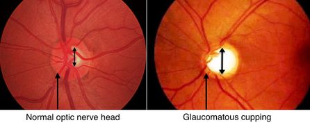
Open-angle glaucoma (cupping)
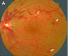
Roth spots due to retinal vein occlusion (retinal hemorrhage)
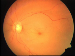
Central retinal artery occlusion: cherry-red spot, retinal edema and narrowing of the vessels.


Other
- Tono-Pen/Applinator/Schiotz
- EOM
- Visual fields
- Xray with concerns of metal/glass/stone
- See here for Optokinetic drum
See Also
References
- ↑ Rupp JD. The 8-Point Eye Exam. American Academy of Ophthalmology. MAY 24, 2016. http://www.aao.org/young-ophthalmologists/yo-info/article/how-to-conduct-eight-point-ophthalmology-exam.