General approach to rashes: Difference between revisions
| (2 intermediate revisions by the same user not shown) | |||
| Line 1: | Line 1: | ||
''This page is for adult patients; for other age groups see [[pediatric rashes]] and [[neonatal rashes]]'' | ''This page is for adult patients; for other age groups see [[pediatric rashes]] and [[neonatal rashes]]'' | ||
==Background== | ==Background== | ||
{{Skin anatomy background images}} | |||
[[File:3D medical animation skin layers.jpg|thumb|3D medical illustration showing major layers of skin]] | [[File:3D medical animation skin layers.jpg|thumb|3D medical illustration showing major layers of skin]] | ||
*A wide range of benign and dangerous pathology can present with a rash | *A wide range of benign and dangerous pathology can present with a rash | ||
| Line 32: | Line 33: | ||
{{DDX dark raised lesions}} | {{DDX dark raised lesions}} | ||
{{Generalized rash DDX}} | {{Generalized rash DDX}} | ||
{{Plaques DDX}} | |||
==Evaluation== | ==Evaluation== | ||
[[File:Dermatologic Emergencies.png|1500px|Algorithm for the Evaluation of Dermatologic Emergencies]] | [[File:Dermatologic Emergencies.png|1500px|Algorithm for the Evaluation of Dermatologic Emergencies]] | ||
{{Maculopapular rashes images}} | {{Maculopapular rashes images}} | ||
{{Bullous rashes images}} | |||
{{Erythematous rashes images}} | {{Erythematous rashes images}} | ||
{{ | {{Purpura rash images}} | ||
{{Dark raised skin lesions images}} | {{Dark raised skin lesions images}} | ||
{{General rashes images}} | |||
==Management== | ==Management== | ||
Latest revision as of 16:35, 11 December 2024
This page is for adult patients; for other age groups see pediatric rashes and neonatal rashes
Background


- A wide range of benign and dangerous pathology can present with a rash
Rash Red Flags[1]
- Fever
- Toxic appearance
- Hypotension
- Mucosal lesions
- Severe pain
- Very old or young age
- Immunosuppressed
- New medication
Dermatology Nomenclature
Small lesions (<0.5cm)
| Name | Raised/Palpable | Fluid-Filled | Other Description | Diagram |
| Macule | No | None | flat, cirumscribed, colored | 
|
| Papule | Yes | None | Solid | 
|
| Vesicle | Yes | Clear | .png)
| |
| Pustule | Yes | Pus | Leukocytes or keratin | 
|
Large lesions (>0.5cm)
| Name | Raised/Palpable | Fluid-Filled | Other Description | Diagram |
| Patch | No | None | Large macule (flat, colored) | 
|
| Plaque | Yes | None | Superficially raised, circumscribed solid area | 
|
| Nodule | Yes | None | Distinct large papule | 
|
| Bulla | Yes | Clear | Large vesicle/blister or exposed epidermal layer | 
|
| Wheal | Yes | Edema | Firm and edema of dermis |
Other

- Eschar
- Fissure/erosion/ulcer
- Necrotizing rashes
Clinical Features
History
- Key elements from the history include:
- Distribution and progression of the skin lesions
- Recent exposures (sick contacts, foreign travel, sexual history and vaccination status)
- Any new medications
Physical Exam
- Pay specific attention to vital signs
- A rash associated with fever or hypotension should make you worry about potentially deadly diagnoses
- Perform a careful physical exam
- Undressing the patient to fully examine the trunk and the extremities
- Look at palms, soles and mucous membranes
- Touch the skin with a gloved hand to determine if the lesions are flat or raised
- Press on lesions to see whether they blanch
- Rub erythematous skin to see if it sloughs
Differential Diagnosis
Maculopapular rashes
- Infectious
- Noninfectious
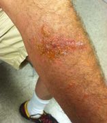
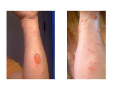
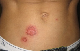
Vesiculobullous rashes
Febrile
- Diffuse distribution
- Varicella (chickenpox)
- Smallpox
- Monkeypox
- Disseminated gonococcal disease
- DIC
- Purpural fulminans
- Localized distribution
Afebrile
- Diffuse distribution
- Bullous pemphigoid
- Drug-Induced bullous disorders
- Pemphigus vulgaris
- Phytophotodermatitis
- Erythema multiforme major
- Bullous impetigo
- Localized distribution
- Contact dermatitis
- Herpes zoster (shingles)
- Dyshidrotic eczema
- Burn
- Dermatitis herpetiformis
- Erythema multiforme minor
- Poison Oak, Ivy, Sumac dermatitis
- Bullosis diabeticorum
- Bullous impetigo
- Folliculitis
Necrotizing rashes
- Necrotizing soft tissue infections
- Purpura fulminans
- Drug rash
- Levamisole toxicity
- Heparin-induced skin necrosis
- Warfarin-induced skin necrosis
Petechiae/Purpura (by cause)
- Abnormal platelet count and/or coagulation
- Septicemia
- Idiopathic thrombocytopenic purpura (ITP)
- Hemolytic uremic syndrome
- Leukemia
- Coagulopathies (e.g. hemophilia)
- Henoch-Schonlein Purpura (HSP)
- Acute hemorrhagic edema of infancy (AHEI)
- Hypersensitivity vasculitis
- Primary vasculitides
- Wegener's
- Microscopic polyangiitis
- Eosinophilic granulomatosis with polyangiitis (Churg-Strauss syndrome)
- Secondary vasculitides
- Trauma
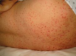
Erythematous rash
- Positive Nikolsky’s sign
- Febrile
- Staphylococcal scalded skin syndrome (children)
- Toxic epidermal necrolysis/SJS (adults)
- Afebrile
- Febrile
- Negative Nikolsky’s sign
- Febrile
- Afebrile
Dark raised skin lesions
Other Rash
- Acute generalized exanthematous pustulosis
- Allergic reaction
- Aphthous stomatitis
- Atopic dermatitis
- Coxsackie
- Dermatitis herpetiformis
- Exfoliative erythroderma
- Impetigo
- Pellagra
- Pityriasis rosea
- Serum Sickness
- Tinea capitus
- Tinea corporis
- Vitiligo
Plaques
- Psoriasis
- Bowen disease
- Discoid lupus erythematosus
- Drug eruption
- Erythema annulare centrifugum
- Lichen planus
- Lichen simplex chronicus
- Nummular dermatitis (nummular eczema)
- Parapsoriasis
- Pityriasis rosea
- Seborrheic dermatitis
Evaluation

Maculopapular rashes visual diagnosis
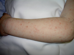
Miliaria (Heat rash)
Poison ivy/Oak/Sumac
Poison ivy/Oak/Sumac


Vesiculobullous rashes visual diagnosis
Bullous impetigo (after the bulla have broken)
Poison ivy/Oak/Sumac
Poison ivy/Oak/Sumac


Erythematous rash


_Skin_Syndrome_-_Feet_Collage.jpg)


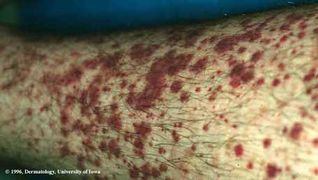
Purpural Rash
Petechiae
Henoch-schonlein purpura (Palpable purpura)
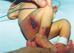
Neonatal purpura fulminans


Dark raised skin lesions


Other Rash visual diagnosis
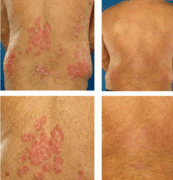
Psoriasis before and after treatment.


Management
- Based on diagnosis
Disposition
- Based on diagnosis
See Also
- Fever and rash
- Pediatric rashes
- Visual diagnosis (main)
- Ulcerative STDs
- Dermatologic nomenclature
- Pigmented rashes
- Rashes of pregnancy
External Links
References
- ↑ Nguyen T and Freedman J. Dermatologic Emergencies: Diagnosing and Managing Life-Threatening Rashes. Emergency Medicine Practice. September 2002 volume 4 no 9.