Periorbital swelling: Difference between revisions
No edit summary |
No edit summary |
||
| Line 4: | Line 4: | ||
[[File:eyelid glands.png|thumb]] | [[File:eyelid glands.png|thumb]] | ||
[[File:Tear system.png|thumb|Right eye lacrimal system consisting of: of lacrimal gland (a), punctums (b,e), canalicules (c,f), lacrimal sac (g,d).]] | [[File:Tear system.png|thumb|Right eye lacrimal system consisting of: of lacrimal gland (a), punctums (b,e), canalicules (c,f), lacrimal sac (g,d).]] | ||
==Clinical Features== | ==Clinical Features== | ||
Revision as of 22:16, 23 October 2024
Background


Clinical Features
Periorbital swelling images

Blepharitis of eyelashes.
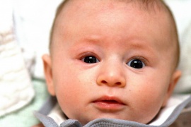
Infant Blepharitis (right)

Bilateral dacryoadenitis

External stye

Internal stye

Acute allergic conjunctivitis

Chronic allergic conjunctivitis

Contact blepharoconjunctivitis

Proptosis due to retrobulbar abscess and orbital cellulitis


Differential Diagnosis
Periorbital swelling
Proptosis
- Normal IOP
- Orbital cellulitis
- Orbital pseudotumor
- Orbital tumor
- Increased IOP
- Retrobulbar abscess
- Retrobulbar emphysema
- Retrobulbar hemorrhage
- Ocular compartment syndrome
- Orbital tumor
No proptosis
- Periorbital cellulitis/erysipelas
- Dacryocystitis (lacrimal duct)
- Dacryocele/Dacryocystocele
- Dacryostenosis
- Dacryoadenitis (lacrimal gland)
- Allergic reaction
- Nephrotic Syndrome (pediatrics)
Lid Complications
- Blepharitis (crusts)
- Chalazion (meibomian gland)
- Stye (hordeolum) (eyelash folicle)
Other
- Subperiosteal abscess
- Orbital abscess
- Cavernous sinus thrombosis
- Conjunctivitis
- Contact dermatitis
- Herpes zoster
- Herpes simplex
- Sarcoidosis
- Granulomatosis with polyangiitis
Evaluation
Management
- Diagnosis dependent
Disposition
- Diagnosis dependent
See Also
Eye Algorithms
- Red eye
- Periorbital swelling
- Acute vision loss (noninflamed)
- Acute onset flashers and floaters
- Painful eyes with normal exam
- Neonatal eye problems